
Presentation
Nausea, vomiting, weight loss, watery diarrhea, brown skin pigmentation.
Patient Data
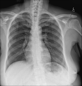
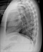

Subtle polypoid nodularity projecting over the stomach bubble in the left upper quadrant. Chest is clear.
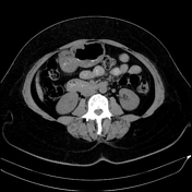

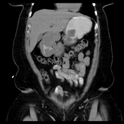

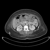
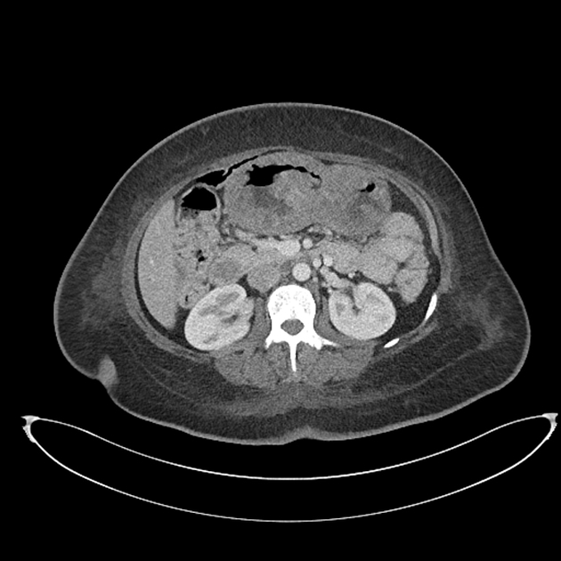
Non-contrast CT with positive oral contrast, which opacifies to the distal small bowel.
Extensive thickening and polypoid nodularity of the stomach and duodenum. Large bowel appears generally unremarkable.
Several peritoneal nodules of uncertain significance.

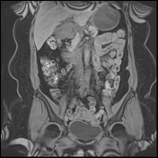
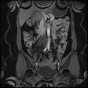
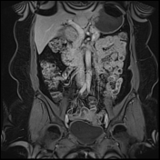
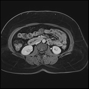
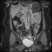
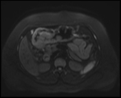
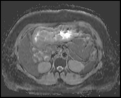
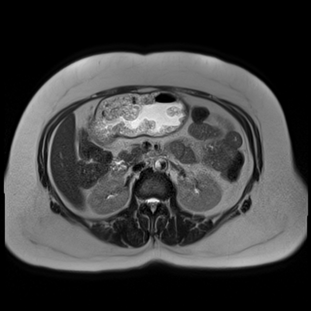
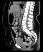
MR ENTEROGRAPHY
There are innumerable polyps in the stomach and first part of duodenum of varying size and morphology, most numerous in the distal body and antrum of the stomach where they are confluent and "carpeting" the gastric mucosa. Sparing of the gastric fundus.
The gastroduodenal polyps demonstrate moderately hyperintense signal and have a smoothly lobulated, frond-like outline with no obvious extension through the gastric wall
Appearances favor a polyposis syndrome, including Peutz-Jeghers, Cowden, Cronkhite-Canada or juvenile polyposis syndrome. Absence of polyps in the fundus makes familial adenomatous polyposis less likely.
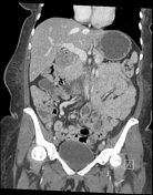
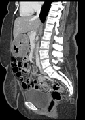
Further CT imaging was performed at 10 months as patient presented with clinical suspicion of septicemia.
Irregular polypoid thickening and edema of the gastric wall (distal body and antrum) and duodenum. These appear more prominent than initial CT, likely reflecting developing edema. Proximal ascending colon wall thickening, and possible thickening of the recto-sigmoid junction.
An ileoileal intussusception is also present within the right upper quadrant.
Case Discussion
Upper GI endoscopy:
Gastro-esophageal junction nodule.
Pedunculated and sessile polyps throughout stomach and carpet-like polyps in duodenum
Colonoscopy:
Hemorrhagic, ulcerated and nodular rectosigmoid and sigmoid colon.
Histology:
Biopsies from GEJ, stomach, duodenum and sigmoid:
All biopsies showed similar histological features, with no evidence of malignancy. Foveolar hyperplasia was present. Specific features were highly suggestive of Cronkhite-Canada, although a rare variant of juvenile polyposis syndrome could not be entirely excluded microscopically.
---
Discussion:
Cronkhite-Canada is a rare hamartomatous polyposis syndrome, affecting the stomach and large bowel, with relative sparing of the esophagus and small bowel. The presence of a GEJ polyp in this case is slightly atypical, but otherwise the clinical features in combination with imaging findings, endoscopy and histology all point to Cronkhite-Canada syndrome as the cause.


 Unable to process the form. Check for errors and try again.