
Presentation
Left flank pain.
Patient Data
Age: 20 years
Gender: Female
From the case:
Crossing vessels causing uretero-pelvic junction obstruction


Show annotations
Download
Info

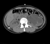
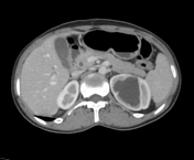
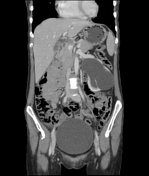
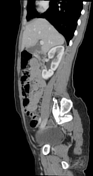
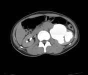
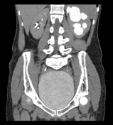
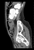
There is evidence that branches of the renal vein cross the pelviureteric junction before that to join in the renal pelvis which is best seen on sagittal view in this case and resulting in severe hydronephrosis and disproportionate dilatation of the renal pelvis.
No radiodense calculus or definite mass lesion is seen.
The right kidney and the rest of the imaged viscera are within normal limits.
Case Discussion
Current CT findings are more in favour of crossing vessels with resultant pelviureteric junction obstruction which is a common scenario as detailed.
Co-contributor Dr, Anwar-ul-Haq Zadran.


 Unable to process the form. Check for errors and try again.