
Presentation
Episodes of vaginal bleeding in postmenopausal patient.
Patient Data
Age: 65 years
Gender: Female
From the case:
Cystic endometrial hyperplasia


Download
Info
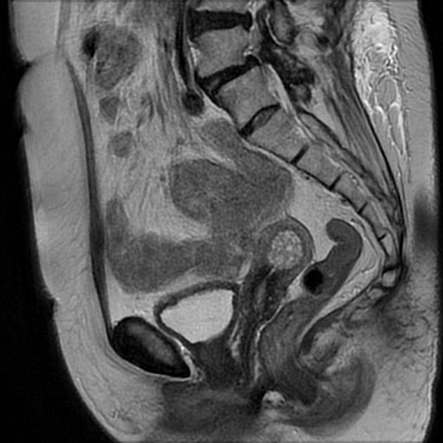
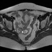
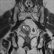
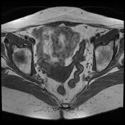
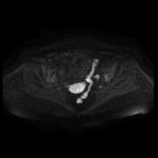
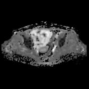
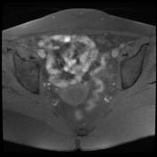
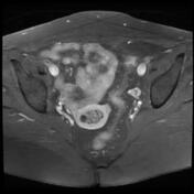
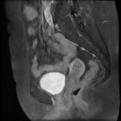
Retroverted retroflexed uterus with a well-defined mass of the endometrial cavity containing numerous tiny cysts of high signal T2 with no restricted diffusion. The postcontrast sequences show moderate heterogeneous enhancement. No underlying myometrial invasion is seen.
Small periurethral cysts (probably Skene duct cysts).
Case Discussion
MRI features a well-defined mass of the endometrial cavity containing numerous tiny cysts with no obvious endometrial invasion (cystic endometrial hyperplasia vs endometrial cancer).
The patient went on to have a total hysterectomy with a histopathological exam that confirms the diagnosis of cystic endometrial hyperplasia.


 Unable to process the form. Check for errors and try again.