Presentation
Palpable mass in the left upper abdomen.
Patient Data
Age: 35 years
Gender: Female
From the case:
Cystic papillary renal cell carcinoma
![](https://prod-images-static.radiopaedia.org/images/1474527/42d43f28fb230c1445b4d434702488_thumb.jpg)
![This study is a stack](/packs/stack-YQKLCKBI.gif)
![](https://prod-images-static.radiopaedia.org/images/1474478/e588c62d14b7801de1505cb59ae61c_thumb.jpg)
![This study is a stack](/packs/stack-YQKLCKBI.gif)
Download
Info
![](https://prod-images-static.radiopaedia.org/images/1474527/42d43f28fb230c1445b4d434702488_big_gallery.jpg)
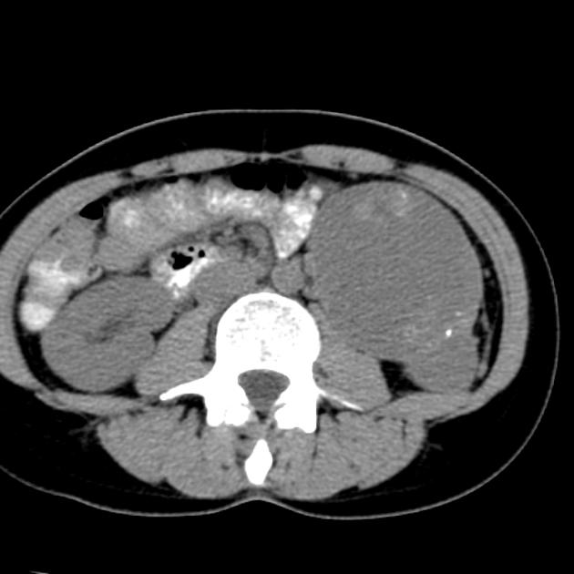
Predominantly cystic mass of the left kidney with enhancing solid calcified nodules.
From the case:
Cystic papillary renal cell carcinoma
![](https://prod-images-static.radiopaedia.org/images/1474630/2ce04ce42b6622f0ab8dd116d7edd1_thumb.jpg)
![This study is a stack](/packs/stack-YQKLCKBI.gif)
![](https://prod-images-static.radiopaedia.org/images/1474694/2e38278802bf04ae2a71718e472e13_thumb.jpg)
![This study is a stack](/packs/stack-YQKLCKBI.gif)
![](https://prod-images-static.radiopaedia.org/images/1474716/b51b10a68ffafd7f65a6aef5159222_thumb.jpg)
![This study is a stack](/packs/stack-YQKLCKBI.gif)
Download
Info
![](https://prod-images-static.radiopaedia.org/images/1474630/2ce04ce42b6622f0ab8dd116d7edd1_big_gallery.jpg)
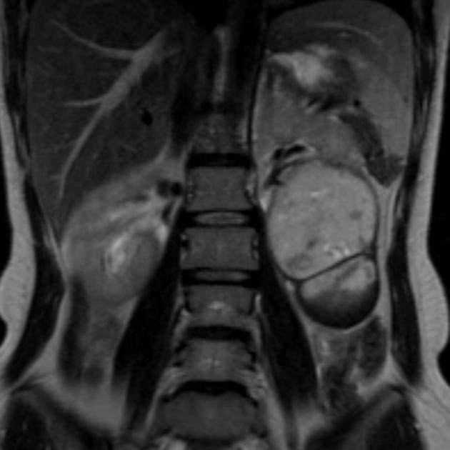
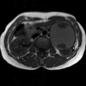
The solid components are more conspicuous on MRI images. A low-signal pseudocapsule and a septation are also present. Moderately hyperintense native T1-signal indicating old hemorrhage.
Case Discussion
Histologic report:
Well-differentiated papillary renal cell carcinoma with regressive changes, hemorrhage and psammom-like calcifications. Focal oncocytic metaplasia.