
Presentation
Left flank pain and hematuria.
Patient Data
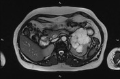

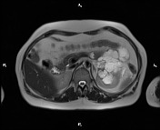
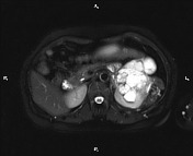
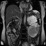
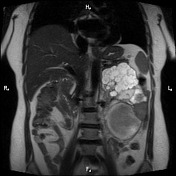
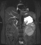
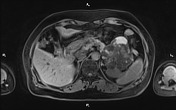
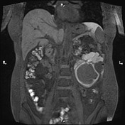
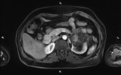
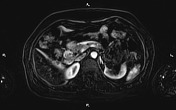
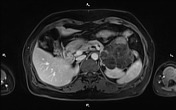
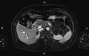
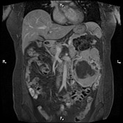
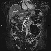
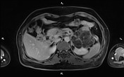
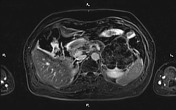
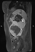
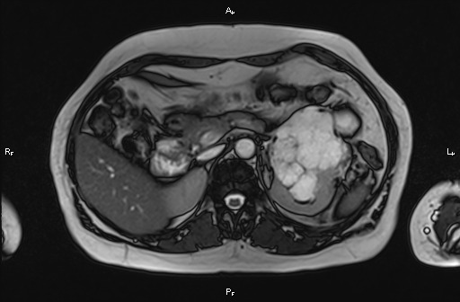
A 122×98×17 mm multilocular cystic mass is seen at lower part of right kidney. Some locules of lesion shows hypersignal contents on T1WI in favor of hemorrhagic contents. There is an enhancing solid component at inferolateral aspect of lesion measuring about 50 mm. There is no sign of vascular extension. The mass causes anteromedial displacement of duodenum and superior displacement of pancreatic tall without any evidences of infiltration. Features are most compatible with Bosniak category 4.
An 18 mm mass is seen at 6th hepatic segment which is iso signal on T1WI and slightly right signal on T2WI. After contrast injection it shows arterial phase hyperenhancement with delayed fading. Benign hyper vascular lesions such as adenoma or FNH are most likely diagnosis.
An 18 mm thin walled cyst is seen at posterosuperior part of spleen.
Case Discussion
The patient underwent left nephrectomy and histopathology evaluation confirms renal cell carcinoma. Additionally US guided core noodle biopsy performed for the hepatic lesion and benign adenoma confirmed.
See also: Bosniak classification of cystic renal masses (version 2019)


 Unable to process the form. Check for errors and try again.