
Decussation of the superior cerebellar peduncle on diffusion tensor imaging
Presentation
N/A
Patient Data
Diffusion tensor imaging (DTI) and tractography in a normal subject depicting the decussation of the superior cerebellar peduncle (SCP).
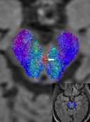
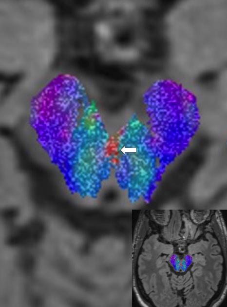
Axial FLAIR MR image with FA (fractional anisotropy) map overlay shows the SCP decussation (arrow, red fibers) centrally in the ventral midbrain.
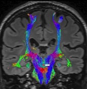
Coronal FLAIR with FA map overlay again depicts the SCP (arrow). By convention, red denotes transversely oriented fibers, blue denotes craniocaudally oriented fibers and green denotes anteroposteriorly oriented fibers on DTI.
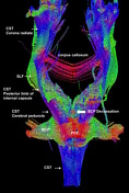
Tractographic reconstructions of the SCP (arrow) in the frontal plane showing the central red dot of the SCP at the level of the midbrain. Abbreviations: CST (corticospinal tract), SLF (superior longitudinal fasciculus), MCP (middle cerebellar peduncle), PCF (pontocerebellar fibers).
Images obtained on a 3 T MRI scanner. Diffusion tensor imaging sequence parameters: diffusion-weighted single-shot spin-echo echo-planar sequence along 30 different geometric directions. An effective b-value of 1000 s/mm2 was used for each of the 30 diffusion-encoding directions. Tractography was performed with the ROI (region of interest) placed in the midbrain. Software setting: tract length 50-400 mm, 2 seed points per voxel length, angle threshold 30 degrees and FA threshold of 0.3.


 Unable to process the form. Check for errors and try again.