
Presentation
One month history of sharp right abdominal and flank pain.
Patient Data


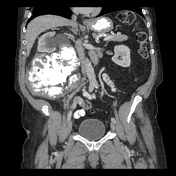
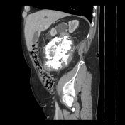
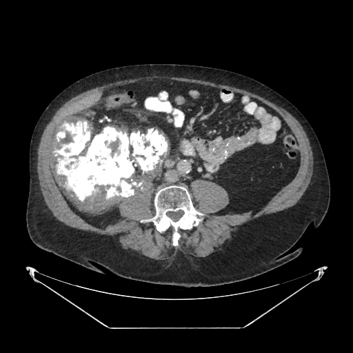
There is a large, heterogenous, partially calcified mass in the right retroperitoneum. The mass exerts a mass effect on the proximal right ureter leading to moderate right hydronephrosis and proximal hydroureter.
Incidental findings:
dilation and tortuosity of the left portal vein, possibly reflecting a portal venous varix
moderate biliary sludge and calcified gallstones in the gallbladder neck
Case Discussion
This is a case of a dedifferentiated retroperitoneal liposarcoma.
The patient underwent a CT-guided core biopsy which revealed mild to moderate cellular proliferation composed of spindled lesional cells with nuclear hyperchromasia and occasional multinucleation in the background of fibrous stroma focally associated with mature-appearing adipose tissue. Mitosis was rare (up to 1 mitoses in 10 high-power fields). No tumor necrosis was seen. Lipoblasts were not identified. In immunohistochemical studies, the lesional cells were positive for SMA (multifocal) and were negative for pancytokeratin, CAM5.2, EMA, CD34, STAT6, S100, SOX10, desmin, MUC4 and ALK1. The DOG1 stain was weakly positive in scattered cells. The fluorescence in-situ hybridization for the MDM2 gene was positive for amplification. The findings are consistent with dedifferentiated liposarcoma, morphologically corresponding to FNCLCC grade 2.


 Unable to process the form. Check for errors and try again.