Presentation
Dyspnea, post prandial abdominal discomfort and regurgitation. Past history of blunt trauma to abdomen in a road traffic accident (RTA).
Patient Data
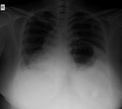
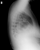
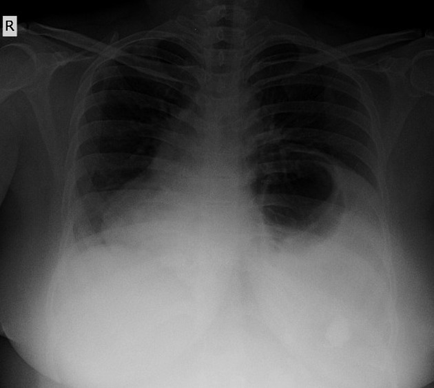
Elevated left hemidiaphragm with abnormally large fundic gas shadow beaneath. There is right sided mediastinal shift. The margin of the left dome of diaphragm cannot be traced completely.


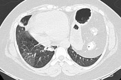

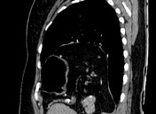

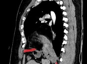

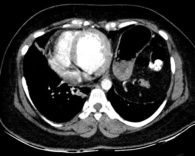
Large sized defect seen in left hemi-diaphragm measuring approximately 10 x 10cm with herniation of the fundus and body of stomach and the splenic flexure of colon through the defect.
Resultant shift of mediastinum towards right with passive atelectasis of the medial segment of the right middle lobe and medial basal segment of the right lower lobe.
Red line represents diaphragm. There is disruption and discontinuity of the red line representing diaphragmatic rupture and herniation through the defect.
Case Discussion
Post- traumatic diaphragmatic hernia is not an uncommon sequel. But lack of awareness of this condition may delay diagnosis and result in life-threatening complications.
CT scan is regarded as the investigative tool of choice but some prefer barium studies in delayed cases of diaphragmatic hernia.
Chest x-ray and sonography of the chest and abdomen may also help in arriving at a diagnosis. An awareness of the condition assisted by the radiological investigations will lead to an early diagnosis and treatment which ultimately helps in managing the patients with diaphragmatic hernias better.


 Unable to process the form. Check for errors and try again.