
Presentation
Leg weakness and foot pain.
Patient Data


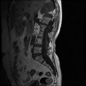
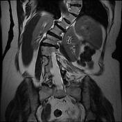
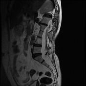
The MRI sequences demonstrate:
marked lumbar scoliosis of left-sided convexity
conus medullaris terminates in a low position at S2 (Tethered cord syndrome)
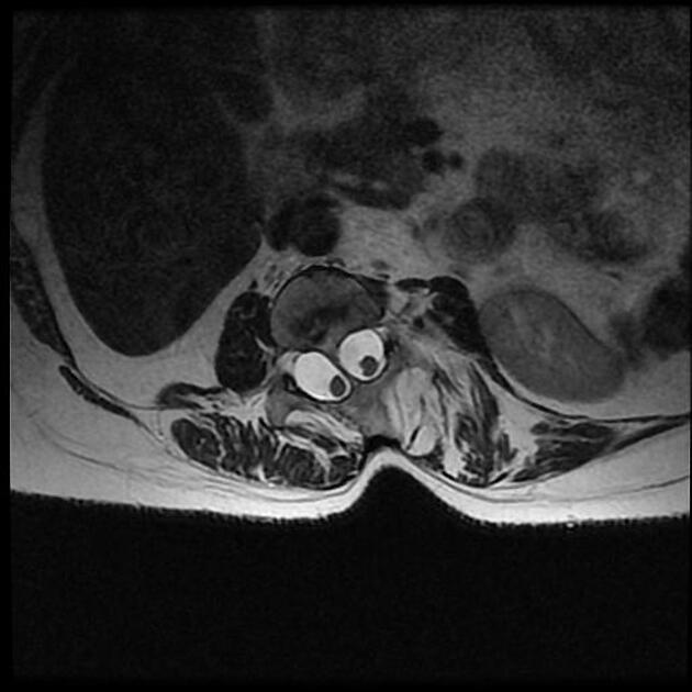
diastematomyelia type I at L2-L3 (hemicords separated by a bony spur)
vertebral abnormalities: butterfly vertebrae of L3 and block vertebrae at L2-L3 and L3-L4 levels
dural ectasia at the lumbar region
spondylotic canal stenosis at D11-D12 level due to disc bulging and ligamentum flavum hypertrophy
Case Discussion
MRI features of diastematomyelia, also known as a split cord malformation type I which is the classic type and characterized by:
duplicated dural sac with midline spur (osseous or osteocartilaginous)
hydromyelia is common
vertebral abnormalities such as hemivertebrae, butterfly vertebrae, spina bifida
cutaneous pigmentation, hypertrichosis, hemangioma are common
patients are usually symptomatic presenting with scoliosis and tethered cord syndrome


 Unable to process the form. Check for errors and try again.