
Presentation
Hemoptysis, not septic.
Patient Data


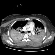
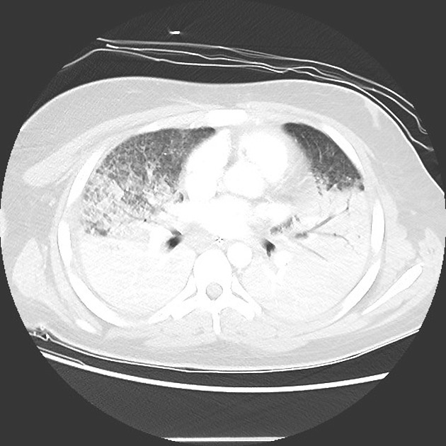
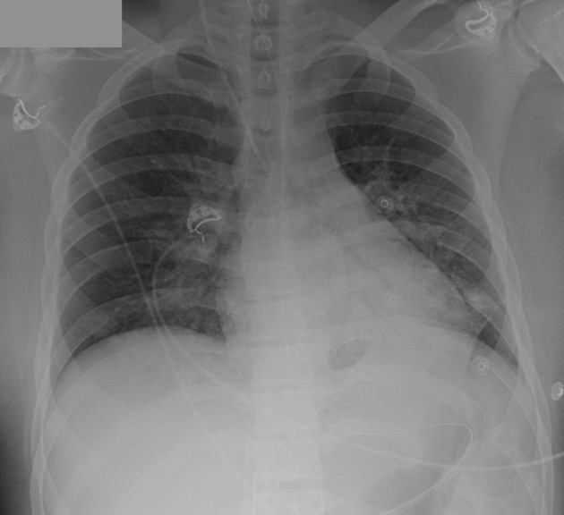
There is widespread lung consolidation permeated by groundglass areas in keeping with alveoli obliteration. The pleural spaces are clear. A few prominent mediastinal lymph nodes are nonspecific.
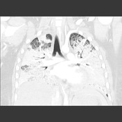
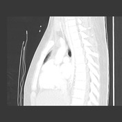
The lungs are mostly clear, with some residual opacities in the perihilar and left lower zones.
Case Discussion
Although non-specific, this diffuse airspace obliteration was thought to be mostly due to alveolar hemorrhage as the presentation was of profuse hemoptysis. No septic markers were elevated to suggest an infective component.
The patient underwent bronchoscopy and biopsy:
Macroscopy: 3 fragments of red-tan tissue, 1-3mm, and multiple <1 mm fragments.
Microscopy: Sections comprise alveolar tissue, within which there is minor pneumocytes hyperplasia, with interstitium, show endothelial hyperplasia and scattered interstitial eosinophils and occasional capillary thrombi. Hemosiderin macrophages are present within the alveolar spaces. Immunofluorescence highlights a linear pattern of IgG deposition along the basement membrane.
Conclusion: Bronchial biopsies, showing features of an active capillaritis, past alveolar bleeding and linear IgG along the basement membrane, diagnostic for Goodpasture's syndrome (anti-GBM antibody disease).
This patient did not have a renal impairment at this time and, although rare, pulmonary or renal involvement may be seen in isolation, mainly in the early manifestation of the disease.


 Unable to process the form. Check for errors and try again.