
Presentation
Seizures
Patient Data


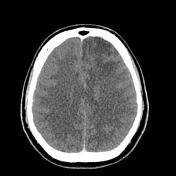
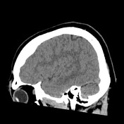
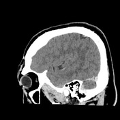
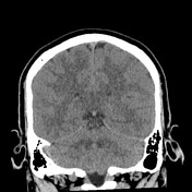
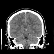
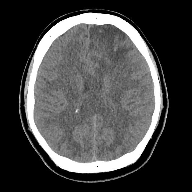
A left frontal region of low density is present with mass effect. A small curvilinear region of increased density is noted, which is of uncertain significance.

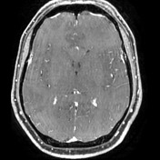

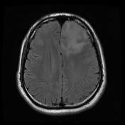
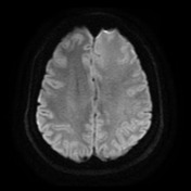

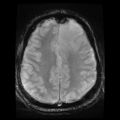
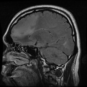
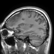
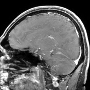
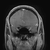
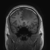
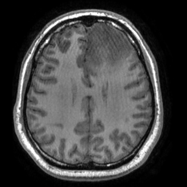
A large left frontal mass is demonstrated without enhancement, calcification, necrosis or hemorrhage.
Features favor a diffuse low grade glioma.
Case Discussion
The patient went on to have surgery.
Histology
The sections show features of a moderately cellular astrocytic tumor. The tumor cells have elongated, angulated and hyperchromatic nuclei. Some gemistocytes are noted, occupying about 5% of the cell population. 1 mitosis is seen. No microvascular proliferation or necrosis is present. There is no oligodendroglial component. The features are those of diffuse astrocytoma.
The tumor cells are p53 and IDH-1 positive (mutated). ATRX shows loss of staining (mutated). MGMT is negative (likely methylated). The topoisomerase index is about 3%.
Final diagnose
Astrocytoma, IDH-mutant (WHO Grade 2)


 Unable to process the form. Check for errors and try again.