
Presentation
This 9-year-old boy was involved in a motor vehicle accident.
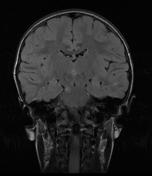
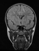
Patient Data

small hyperdense foci of hemorrhage in the left frontal lobe
Due to persistent reduced consciousness, the patient was referred for MRI.
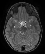
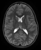
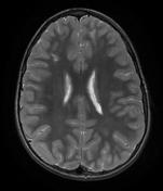
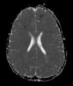
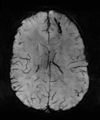
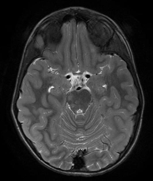
Right-sided focal areas of hyperintense T2 signal in the brainstem, splenium of the corpus callosum, superior frontal gyrus on the left and middle frontal gyrus on the right.
Focal areas of hyperintense FLAIR signal in the splenium of the corpus callosum as well as the periventricular white matter, in the brainstem on the right and subcortical in the middle temporal lobe on the left, in the superior frontal gyrus on the left and middle frontal gyrus on the right.
Focal areas of restricted diffusion in the frontal white matter and splenium of the corpus callosum.
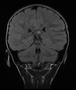
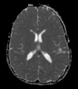
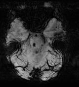
Focal area of signal loss on SWI sequences in the superior frontal gyrus on the left, as well as multiple punctate areas of signal loss bilaterally, indicating hemorrhagic lesions.
Case Discussion
This is a case of diffuse axonal injuries in classic locations:
- gray/white matter interface (predominantly in frontal and temporal lobes)
- corpus callosum (especially the splenium)
- dorsolateral midbrain
The lesions appear as T2/FLAIR hyperintense foci which show restricted diffusion. Hemorrhagic lesions additionally show hypointense signal in T2* gradient echo and susceptibility weighted images (SWI).


 Unable to process the form. Check for errors and try again.