
Presentation
Bilateral inguino-scrotal hernia past 2 years. Abdominal distension past 6 months
Patient Data
Age: 60 years
Gender: Male
From the case:
Diffuse intrabdominal lipomatosis
Download
Info
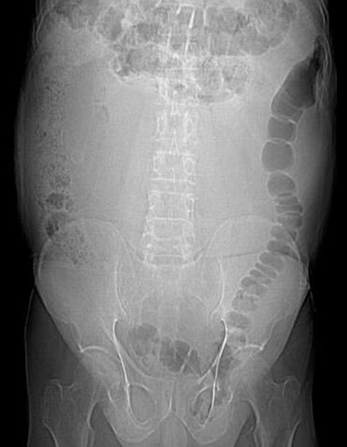
The bowel loops are arranged peripherally no central gas shadow is seen.
From the case:
Diffuse intrabdominal lipomatosis


Download
Info
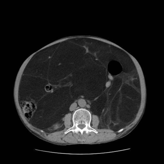
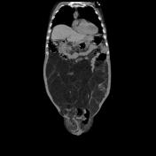
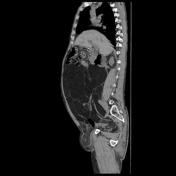
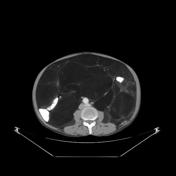
On CT the bowel loops are seen peripherally (superiorly and laterally) in the abdominal cavity.
Large lobulated area of average HU -104 (fat) is seen filling the abdominal and pelvic cavity, displacing the abdominal organs.
Bilateral inguino-scrotal hernias are noted.
From the case:
Diffuse intrabdominal lipomatosis
Download
Info
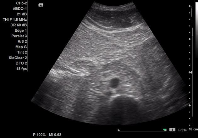
Abdominal and cavity is filled with echogenic and septated amorphous mass which is displacing the abdominal organs.
Case Discussion
Intraperitoneal lipomatosis uncommon and is presumably related to mediastinal lipomatosis or retroperitoneal and pelvic lipomatosis.


 Unable to process the form. Check for errors and try again.