Presentation
Clinician undertaken ultrasound reports bilateral ovarian cysts and posterior wall fibroids. For surgical planning.
Patient Data
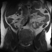

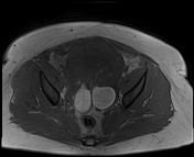
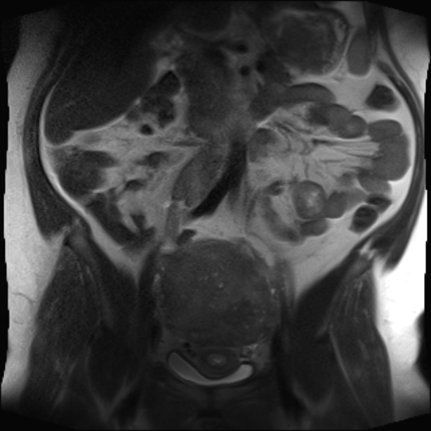
Enlarged uterus. The posterior body is markedly thickened with multiple small high T2 foci within.
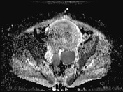
2.4 cm submucosal lesion in the posterior wall of mid body of the uterus indenting the endometrial cavity.
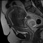
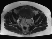
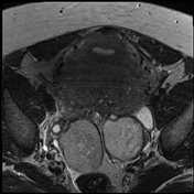
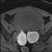
Bilateral high T1, T2 surfaced shaded ovarian cysts measuring 6 cm on the left and 5.4 cm on the right.
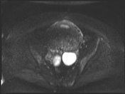
2 cm simple left ovarian cyst.
Case Discussion
Uterine adenomyosis is a condition of menstruating females in which ectopic endometrial tissue is present in the myometrium. It may be diffuse or focal in nature. Given that the condition occurs due to the abnormal siting of endometrial tissue it is often thought of in a similar manner to endometriosis. In fact in 20% of cases, as in this case, is associated with co-existent endometriosis.
Adenomyosis is usually generalized involving large parts of the uterus, typically the posterior wall, but sparing the cervix, as in this case. Although the uterus is enlarged, the overall contour is usually preserved, which is different from the majority of fibroid uteruses.
Endometriomas are focal forms of endometriosis, with the commonest site being in the ovary. They are most frequently unilocular and contain degenerated blood products following repeated cyclical hemorrhage. This is responsible for the characteristic high T1 signal appearance and T2 surface shading appearances.


 Unable to process the form. Check for errors and try again.