
Diffusion tensor imaging and tractography of the cisternal and intradural segment of the trigeminal nerve
Diagnosis not applicable
Patient Data
Gender: Male
MRI Tractography

Download
Info
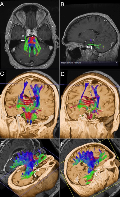
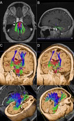
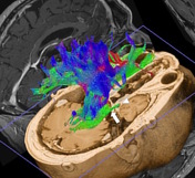
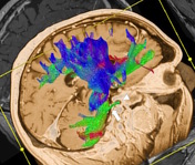
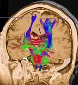
Figure A and B showing diffusion tensor imaging (DTI) with anatomical overly on gadolinium enhanced MPRAGE depict the cisternal (arrow) and intradural (arrow head) segments of the trigeminal nerve in green color. Figure C-F: Tractography showing the normal pathway of the cisternal segment of the trigeminal nerve (arrow) coursing through the pre-pontine cistern into the Meckel's cave.
The 3-D orientation of the major eigenvector was color coded per voxel according to convention with red indicating the transverse (x-element), green indicating the anteroposterior (y-element), and blue indicating the craniocaudal (z-element) orientation.
Post processing performed on the syngo. MR Neuro 3D software.


 Unable to process the form. Check for errors and try again.