
Presentation
History of surgical excision of left cervical cystic lesion, pathologically proved metastatic papillary carcinoma.
Patient Data


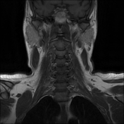
No residual/recurrent lesions at the operative site.
No pathological nodal enlargement.
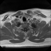
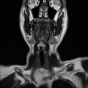
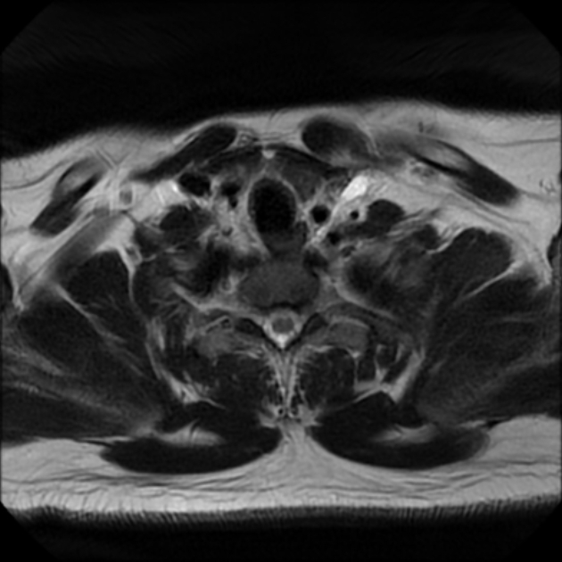
The thyroid gland is slightly of diffuse low signal, yet with no sizable cystic or solid masses.
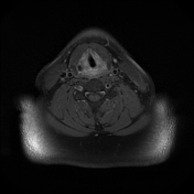
Mild edema signal of the left lower sternomastoid muscle, post-operative sequel.
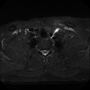
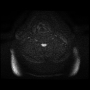
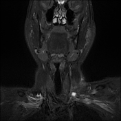
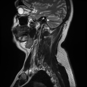
The distal end of the thoracic duct could be visualized and should not be mistaken for a pathology.
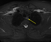
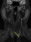
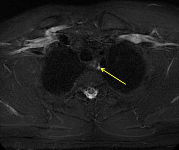
Arrows follow the course of the thoracic duct starting in the mediastinum, tracks upwards, passes behind the left common caarotid arery to end into the left subclavian vein, with ampullarry dilatation of its distal end.
Case Discussion
In this case the patient had a history of cystic lymph node on the left side, which was excised and proven to be metastatic papillary cancer. The left lower cervical tubular cystic lesion was mis-diagnosed by the junior radiologist as a residual cystic necrotic node. However if you track this tubular structure you can see it extending into the left side of the mediastinum. This structure is the terminal end of the thoracic duct.
The thoracic duct is the common trunk for most of the lymphatic vessels that convey lymph into the vascular system. It ends into the left subclavian vein.
The distal thoracic duct can appear on neck CT or MRI.
The thoracic duct typically course within the left side of the neck.The distal thoracic duct usually has a tubular configuration.
Radiologists should be familiar with the normal CT/MRI appearance of the distal thoracic duct to differentiate it from pathologic lesions of the lower neck, such as reactive or metastatic nodes or congenital cysts.


 Unable to process the form. Check for errors and try again.