
Presentation
Patient referred for mammography exam, without additional complaints.
Patient Data
Age: 50 years
Gender: Female
From the case:
Ductal carcinoma in situ
Download
Info
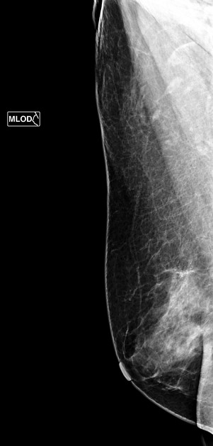
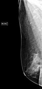
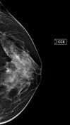
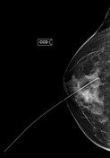
Sparse fibroglandular breasts with pleomorphic microcalcifications grouped at the union of the upper quadrants of the right breast.
From the case:
Ductal carcinoma in situ
Download
Info

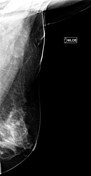
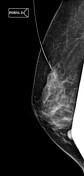
Stereotaxic marking procedure before segmentectomy.
Download
Info
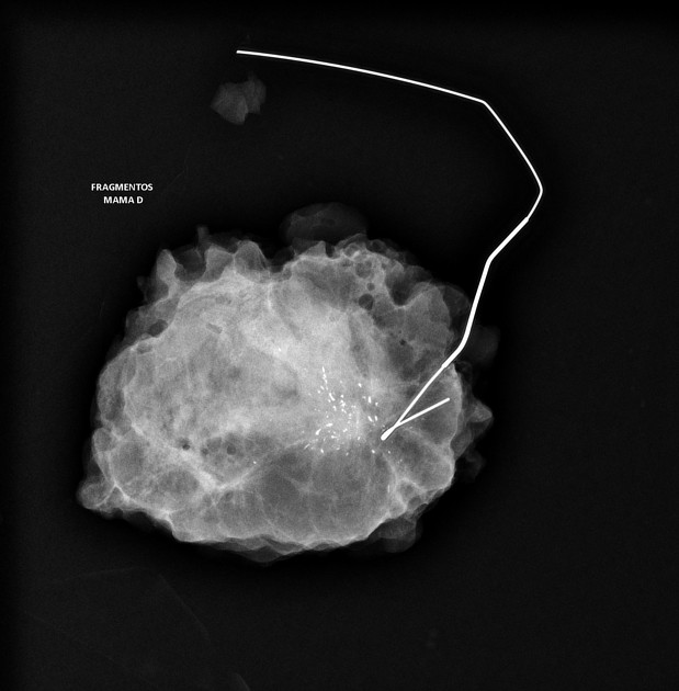
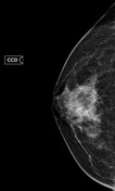
Radiographic image of the resected anatomical specimen showing pleomorphic microcalcifications inside.
Case Discussion
The anatomical specimen obtained from segmentectomy was submitted for anatomopathological evaluation, confirming the diagnosis of ductal carcinoma in situ.
Co-authors: Dr. Hemilianna Hadassa Silva Matozinho M.D. and Dr. Ana Karina de Ataíde Feitosa M.D.


 Unable to process the form. Check for errors and try again.