
Presentation
The patient experienced a fall from about one meter onto a hard surface (left upper abdomen).
Patient Data
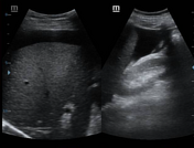
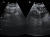
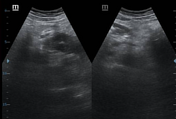
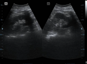
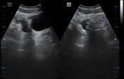
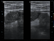
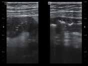
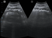
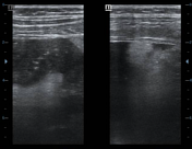
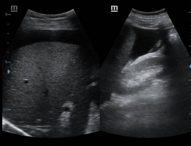
Mild free fluid collection (ascites seen around the liver, spleen, Morison pouch, right para-colic gutter, mildly in the pelvic cavity -200-300ml. Echogenic fat at the upper abdomen, diffuse hypoechoic entire duodenal walls, part of jejunum, consisting of duodenal hematoma. Normal pancreas, spleen, liver kidneys, normal gall bladder, and biliary tree. Normal urinary bladder, no pleural effusion sample of serous fluid was sent for investigation.
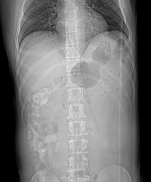
No signs of pneumoperitoneum; trivial gas in the stomach, left upper abdomen, and right center of the abdomen. No dilated bowels observed.
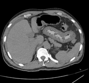

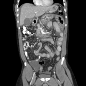
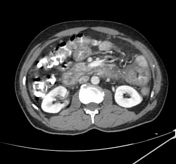
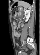

There is diffuse thickening of the walls in the duodenal and long segment of proximal jejunal bowel loops, which may indicate the presence of an intramural hematoma rather than a bowel contusion. Diverticulosis is noted in the transverse colon and proximal descending colon.
There is a mild collection of low-density fluid in the perihepatic and pelvic regions, but no obvious injuries to solid organs are observed. The liver is of average size with a homogeneous density and shows no abnormal densities or dilatation of the intrahepatic biliary tree. The gallbladder and common bile duct appear normal, as do the pancreas and spleen.
Case Discussion
The patient improved under observation. The patient had a classic long segment small bowel wall hematoma involving the duodenum and jejunum. There was no leakage of oral contrast, and aspiration confirmed that the patient was not hemorrhaging.


 Unable to process the form. Check for errors and try again.