
Presentation
Study prompted by ultrasound findings.
Patient Data


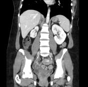
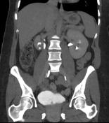
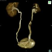

CT shows a left kidney normal in size, shape and cortical thickness. The right kidney is hypoplastic and in the excretory phase shows two separate renal pelvis each with a ureter. There is dilatation of the right distal ureter, and a contrast-filled structure protruding into the lumen of the bladder on the excretory phase, consistent with a ureterocele.
Case Discussion
A ureterocele is a dilation of the distal end of the ureter. This appearance has been called cobra head sign. Ureteroceles may occur in cases of simple or duplicated ureters like in this case. There are two types of ureterocele: orthotopic and ectopic. In an orthotopic ureterocele, the hole in the ureter and the ureterocele is intravesical. In the case of ectopic ureterocele, the ureterocele lies in the submucosa of the bladder and part of ureterocele extends into the bladder neck or urethra.
The orthotopic ureterocele contain one orifice which opens in the normal range but are stenotic. Most of the findings are orthotopic ureterocele. However when they are large, the orthotopic ureterocele may obstruct the bladder neck or cause hydronephrosis. The incidence of persistent infections and stones are increased in both types of ureteroceles.


 Unable to process the form. Check for errors and try again.