
- Note: This case has been tagged as "legacy" as it no longer meets image preparation and/or other case publication guidelines.
From the case:
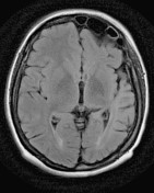
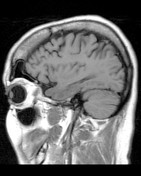
Dyke-Davidoff-Masson syndrome

Download
Info

Selected images from an MRI demonstrates asymmetry of the cerebral hemispheres, with the left appearing atrophic. This is associated with enlargement of the left frontal sinus and thickening of the skull vault (note how much more marrow can be seen on the left). Findings are consistent with Dyke-Davidoff-Masson Syndrome.


 Unable to process the form. Check for errors and try again.