
Presentation
Dysphagia
Patient Data
Age: 60
Gender: Female
From the case:
Dysphagia lusoria

Download
Info

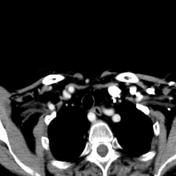
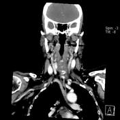
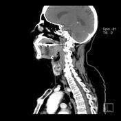
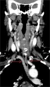
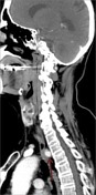
Left sided aortic arch with aberrant right subclavian artery is seen extrinsically indenting the posterior wall of the upper thoracic esophagus; it's also mildly indented by tortuous proximal left common carotid artery crossing along its left lateral aspect.
Asymmetric mild enlargement of both lingual & palatine tonsils, more on the right.
From the case:
Dysphagia lusoria
Download
Info
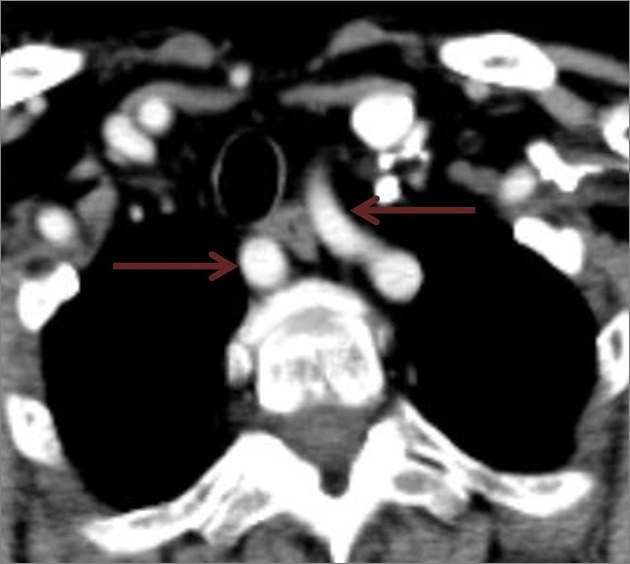
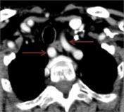
Left sided aortic arch with aberrant right subclavian artery is seen extrinsically indenting the posterior wall of the upper thoracic esophagus; it's also mildly indented by tortuous proximal left common carotid artery crossing along its left lateral aspect. (Arrows)


 Unable to process the form. Check for errors and try again.