
Patient Data
Age: 50 years
Gender: Female


Download
Info
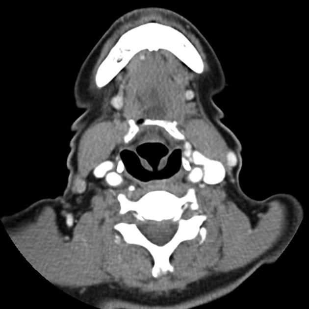
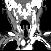
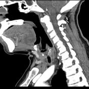
Note the abnormally shaped and small thyroid with a similar nodule of tissue posterior to the esophagus.
Case Discussion
No history of surgery or carcinoma.
These CT findings were confirmed to be thyroid tissue on a thyroid scan, thus representing ectopic thyroid. The patient was followed and no change detected.
Case courtesy of Bob Cook, MD. Western Memorial Regional Hospital Corner Brook, Newfoundland.


 Unable to process the form. Check for errors and try again.