
Patient Data
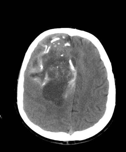
A large mass with prominent calcification and mass effect.
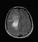

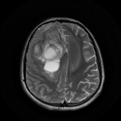
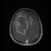
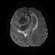
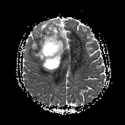
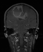
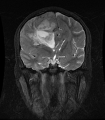

A very large heterogeneous mass is centered on the right frontal lobe with prominent extension across the corpus callosum. Multiple areas of non-enhancement suggest necrosis.
Case Discussion
A young male patient with pathologically proven sPNET.
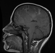
Note: The age of the patient has not been provided, but looking at the dentition it appears to be a young adult. The diagnosis of supratentorial PNET/embryonal tumors with multilayered rosettes (ETMR) is rare outside of the first decade. As the saying goes "extraordinary claims require extraordinary proof" and as such, without more supporting evidence, the diagnostic certainty has been reduced to "possible".
In an adult, it is probably more likely that this represents as glioblastoma with primitive neuronal component, possibly arising from an underlying oligodendroglioma (given the calcifications).
Note: The current (2016) WHO classification of CNS tumors has made substantial changes to tumors previously considered to be supratentorial PNET, now classified as embryonal tumors with multilayered rosettes (ETMR), along with a number of other entities, in recognition of characteristic amplification of the C19MC region on chromosome 19 (19q13.42).


 Unable to process the form. Check for errors and try again.