Presentation
An elderly diabetic gentleman presented to the ED with fever and abdominal pain. On examination, there was diffuse tenderness and guarding. White cell count and C reactive protein were elevated.
Patient Data


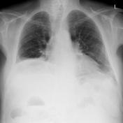
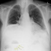
Erect chest x-ray done to look for free gas under the diaphragm.
There is no free gas under the diaphragm. In the right upper quadrant, there is an air-fluid level with a curvilinear lucency visible around the fluid component (see Series 2, annotated).

The patient is getting progressively sicker.
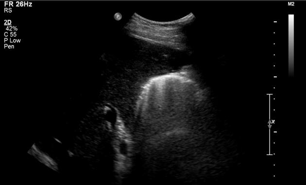
Ultrasound demonstrates a distended gallbladder with an ill-defined wall and dirty shadowing, indicating intramural gas, and in keeping with emphysematous cholecystitis.

Contrast-enhaned CT abdomen confirms a gallbladder distended with gas and fluid, and intramural gas.
Case Discussion
The radiographic appearances of the gas-filled gallbladder with intramural gas are quite different from normal gas-filled structures, e.g. the adjacent hepatic flexure. The differential diagnosis for curvilinear lucency as seen in this case is pneumatosis.
On ultrasound, intramural gas of emphysematous cholecystitis produced dirty shadowing. The differential diagnosis is porcelain gallbladder, where shadowing is usually dense.


 Unable to process the form. Check for errors and try again.