
From the case:
Empyema


Download
Info
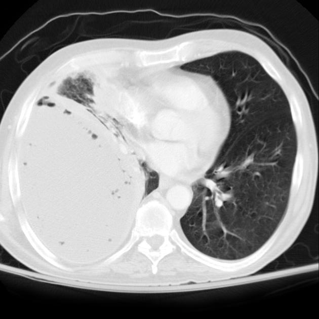
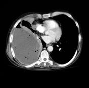
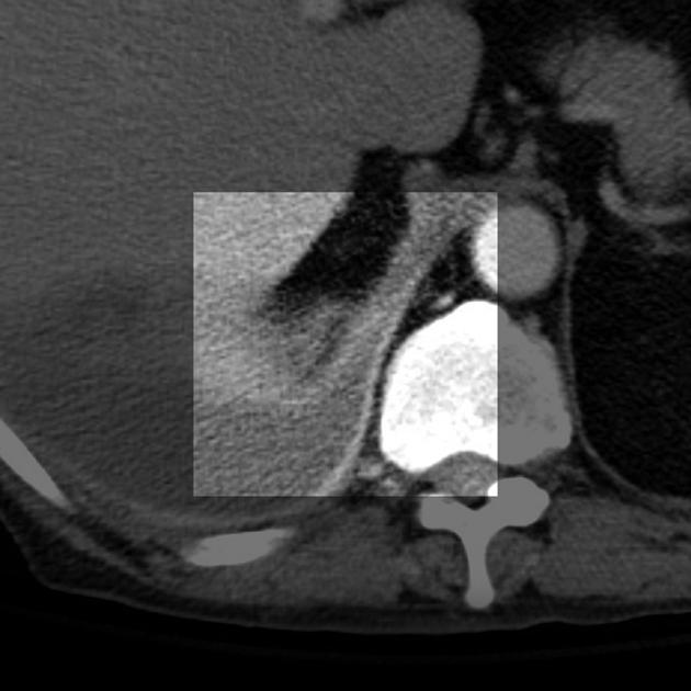
CT of the chest demonstrates a very large right sided pleural collection with thickened surrounding pleura (the so called split pleura sign) and multiple gas bubbles. The adjacent lung is compressed and collapsed.
The findings are consistent with an empyema.
From the case:
Empyema
Download
Info
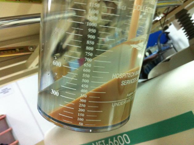
Pleural drain content from an empyema. Note: this image is from a different patient, but with identical clinical features and diagnosis.
Photo credit: Dr Ian Bickle


 Unable to process the form. Check for errors and try again.