
Presentation
Painful left middle finger.
Patient Data

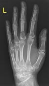
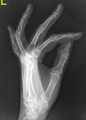
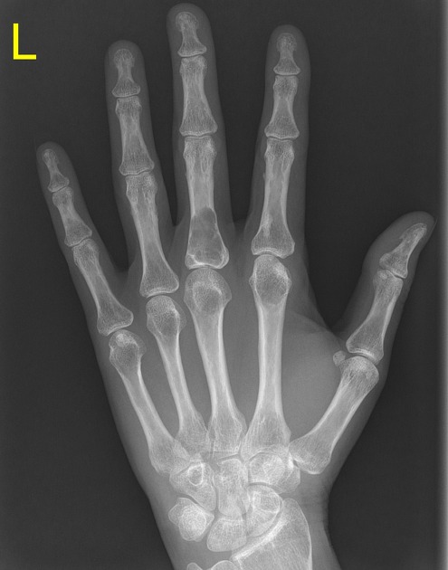
There is a lytic expansile and mildly lobulated lesion with evidence of internal matrix centered within the base of the proximal phalanx. Associated surrounding cortical thinning. No evidence of an acute fracture.
This most likely represents an enchondroma. Other differentials for this appearance would include a giant cell tumor or an aneurysmal bone cyst (with ABC thought much less liklely).
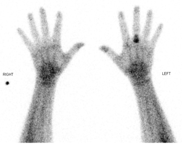
On delayed planar imaging, intense osteoblastic activity is seen within the proximal phalanx of the left 3rd finger, consistent with an enchondroma.
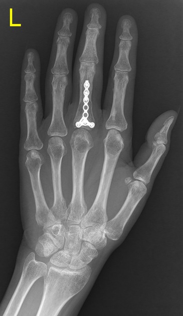
Plate and screw fixation across 3rd proximal phalanx enchondroma.
Histopathology report
The sections show large fragments of hypocellular cartilaginous tissue. There is no myxoid change seen. There is no atypia and the nuclei are small.
The histological features are likely those of an enchondroma, with the appropriate radiological correlation.
Case Discussion
Key learning points:
- enchondromas are small, benign lytic bone lesions
- can still result in pathological fractures (with prophylactic internal fixation in this case)
- increased uptake on bone scan can be seen with enchondromas particularly with underlying pathological fracture or cortical expansion in small bones


 Unable to process the form. Check for errors and try again.