
Presentation
Painful left knee.
Patient Data
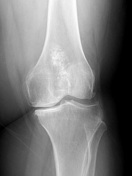
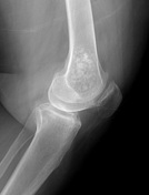

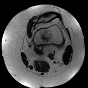
Moderate osteoarthritic changes are noted, seen as narrowed medial patellofemoral compartment, marginal osteophytes, and eburnated medial tibial plateau.
Lower femur ill-defined intramedullary lesion with chondroid matrix calcifications.
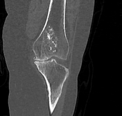

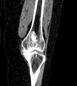

The lower femoral intramedullary lesion clearly depicts chondroid matrix calcifications.
Knee joint osteoarthritic changes are noted.

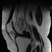
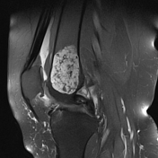
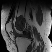
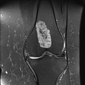
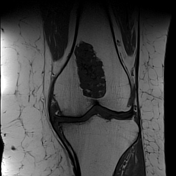
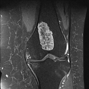
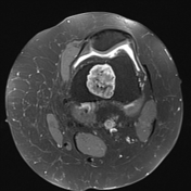
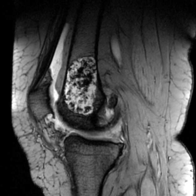
A large well defined central medullary focal bony lesion is noted at the distal left femoral shaft, with the following features:
- it measures 3.1 x 2.7 x 5.2 cm in maximum dimensions
- it expresses heterogeneous T1 hypo and T2 hyperintense signal with intra-lesional T1 mixed hyper and hypointense and T2 hypointense foci
- no diffusion restriction in DWI
- it showed moderate heterogeneous post-contrast enhancement
- it showed lobulated outlines with marrow zone of transition and no surrounding marrow signal abnormalities
- intact overlying cortex
- no related periosteal reaction
- no extension to the nearby soft tissues
Also noted:
- thinned out retropatellar and medial femorotibial cartilage, with subchondral edematous marrow changes as well as marginal osteophytes noted, reflecting osteoarthrosis
- bulky lateral meniscus impending discoid formation showing transverse linear T2 hyperintense signal noted within its body reaching its superior articular surface reflecting horizontal tear
- radial tear at posterior horn medial meniscus interrupting its superior and inferior articular surfaces
- mild knee joint effusion
- thin T2 hyperintense signal noted superficial to the infrapatellar tendon, reflecting mild bursitis
Case Discussion
Overall features matching with a sizable distal femoral non-aggressive chondrogenic lesion, suggestive of a low-grade chondral lesion, almost certainly an enchondroma. Enchondroma is a benign cartilaginous lesion of the medullary cavity. Most enchondromas are asymptomatic and incidentally noted.
Other findings include, impending discoid formation of lateral meniscus showing horizontal tear at its body, radial tear at posterior horn medial meniscus, patellofemoral and medial femorotibial osteoarthrosis and infrapatellar bursitis.
Case courtesy Dr Rim Bastawy, Lecturer of radiodiagnosis, Alexandria University, Egypt.


 Unable to process the form. Check for errors and try again.