
Patient Data
Age: 60 years
Gender: Female
From the case:
Endometrial carcinoma


Download
Info
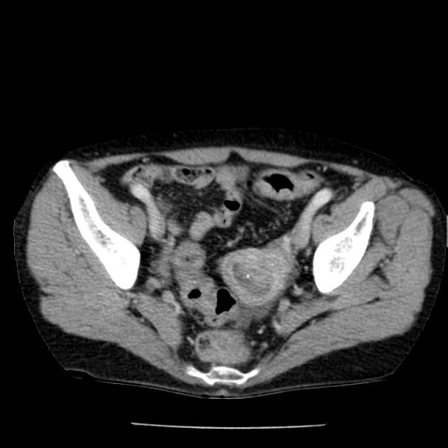
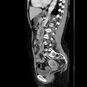
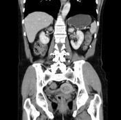
There is a thickened uterus wall with a round configuration., with a small punctate calcification that protrudes into the uterine cavity. The endometrial cavity is dilated and heterogeneously hypodense. There is a punctate calcification within the endometrial cavity.
Case Discussion
The tomographic findings suggest an endometrial cancer. The biopsy proved to be an adenocarcinoma of the endometrium.


 Unable to process the form. Check for errors and try again.