
Presentation
Left adnexal lesion on ultrasound
Patient Data
Age: 25 years
Gender: Female


Download
Info
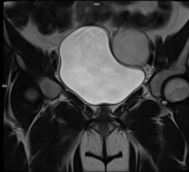
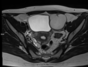
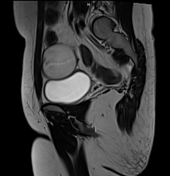
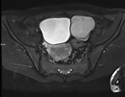
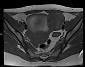
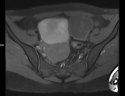
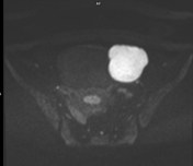
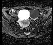
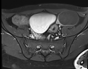
The well-defined lesion in the left ovary. The lesion appears intermediate signal intensity on T2WI, isointense on T1WI, diffusion restriction on DWI and thin peripheral rim enhancement on T1W fat sat post-contrast images with no evidence of signal suppression on T1W/T2W fat-sat images. Tiny hypointense focus within the lesion on T2WI (T2 dark spot sign).
Case Discussion
The MRI findings are most likely compatible with endometrioma.


 Unable to process the form. Check for errors and try again.