
Presentation
Admitted with seizures and reduced GCS. CXR to confirm ETT position.
Patient Data

Supine radiograph.
ETT tip projects over the right main bronchus. There is corresponding loss of the left hemidiaphragm, with volume loss and mediastinal shift to the left.
The lungs are otherwise clear.
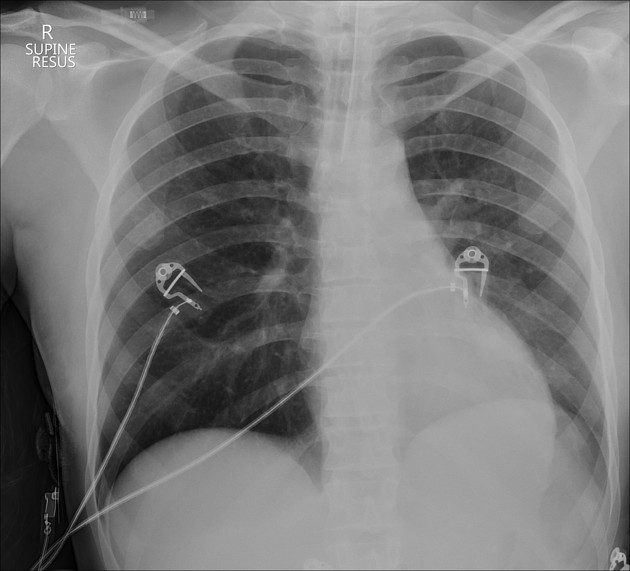
ETT retracted slightly approximately 20 minute interval
Compared with the earlier study, the ETT is now appropriately positioned approximately 2 cm above the carina. Re-expansion of the left lung - the diaphragm is now more visible. The mediastinal shift is also improving (the right cardiac shadow is now to the right of the vertebral bodies).
Case Discussion
This patient's ET tube is initially demonstrated down the right main bronchus, with subsequent left sided collapse.
This was recognized by the clinical team and fixed, with the second CXR showing better positioning and re-expansion of the left lung. This was only over a 20 minute interval and shows how quickly these changes can occur, and the importance of recognizing tube and line positioning on acute studies.


 Unable to process the form. Check for errors and try again.