
Presentation
Patient presented to his GP with 10kg unintentional weight loss. No abdominal pain or localizing symptoms.
Patient Data


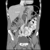

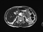
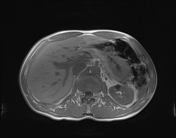
A rounded, fluid-density (~20HU) retrocrural structure is located between the aorta and IVC. It measures 24 x 30 mm. The structure displaces both vessels however fat planes are preserved. There is no solid component or internal complexity and the lesion has a benign apperance.
Differential diagnoses for this finding included an enlarged cisterna chyli, foregut duplication cyst or less likely a cystic GIST.
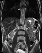
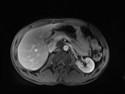

The saccular structure follows fluid signal on all sequences, demonstrating homogeneous T1 hypointensity and T2 hyperintensity. It is continuous with a left lumbar lymphatic trunk and continues superiorly as the thoracic duct.
Case Discussion
The cisterna chyli is formed as the confluence of the lumbar and intra-abdominal lymphatic trunks. It is positioned immediately to the right of the aorta at the L1/2 level.
In one study, average size was measured at 12.9mm (range = 11 to 19 mm).1


 Unable to process the form. Check for errors and try again.