
Presentation
Dizziness.
Patient Data


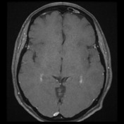
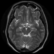
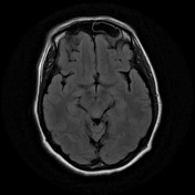

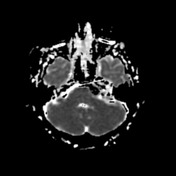
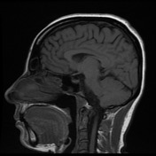
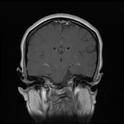
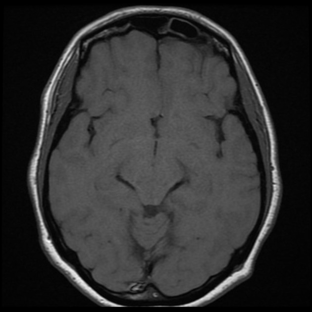
Left cerebellar pontine angle mass extends inferiorly anterolateral to the pons and medulla, to the level of the foramen magnum and superiorly to the tentorial incisura adjacent to the left superior colliculus. Mass characteristics are similar to previous and in keeping with epidermoid. Mild expansion of Meckel's cave on the left, but with no diffusion restriction here. Mild mass effect with compression on the midbrain and left cerebellar peduncle, and displacement of cranial nerve roots through the cerebellar pontine angle similar to previous. No hydrocephalus. Minor mucosal thickening and enhancement of paranasal sinuses, with polypoid mucosal thickening / retention cyst in the left maxillary sinus. Mastoid air cells are clear. Major intracranial flow voids are normal, including major dural venous sinuses. The left transverse sinus is patent but non-dominant.
Case Discussion
The patient went on to have a resection.
Histology
MICROSCOPIC DESCRIPTION: The section shows a collapsed cyst. This is lined by squamous epithelium with a discernible granular layer. The contents comprise laminated keratin. No appendageal structures are identified. The features are of an epidermal cyst. No evidence of tumor is seen. The specimen includes fragments of viable lamellar bone.
FINAL DIAGNOSIS: Epidermoid cyst
Discussion
These are typical findings of an epidermoid cyst, a potentially tricky diagnosis if DWI not available.


 Unable to process the form. Check for errors and try again.