
Presentation
Gradually increasing vertigo, gait disturbance and headaches.
Patient Data


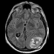
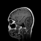
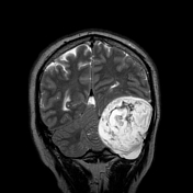
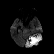
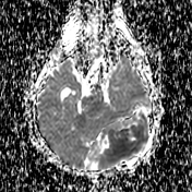

Ovoid mass measuring 77 x 75 x 50mm (FH x RL x AP) located at the left side of posterior cranial fossa with moderate mass effect on the cerebellum, pons, medulla oblongata with compression of 4th ventricle, and the left temporal lobe. Also well seen intra- and transdiploic involvement, but no further galea aponeurotica. There is 6mm of contralateral midline shift and 10mm tonsillar herniation.
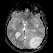
The mass demonstrates heterogeneous T1 and T2 signal intensities, but more valuable is high DWI signal within the mass pronounced centrally, reflecting diffusion restriction, with corresponding low ADC values. There is no significant magnetic susceptibility artifact on gradient echo sequence.
Case Discussion
These MRI findings are in keeping with an intracranial epidermoid cyst.
This patient has a large intracranial epidermoid cyst, which is a congenital lesion with an estimated prevalence about 1% of all intracranial tumors. This lesion was symptomatic and causes mass effect mainly on the cerebellum. It is difficult not to underestimate the value of DWIs - these are short but helpful sequences, especially in identifying epidermoids. So, the use of contrast-enhanced study in this case was doubtful.


 Unable to process the form. Check for errors and try again.