
Presentation
Shortness of breath.
Patient Data

Features are those of a moderate sized left pleural effusion. Bilateral calcified pleural plaques are suggestive of asbestos exposure, the right pleural space is clear. The heart silhouette and the left mediastinal contours are obscured.


Left hemithorax diffuse soft tissue masslike pleural thickening associated with a moderate sized pleural effusion. The pleural masses are better appreciated along the mediastinal margins. Calcified plural plaques again showed. Lungs and airways are otherwise unremarkable. Right hilar prominent lymph node is suspicious for metastasis.
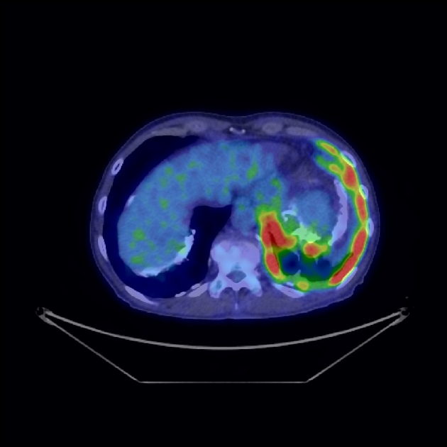
PET-CT demonstrates increased radiotracer uptake along the pleural masses on the left and also within the right hilar and subcarinal lymph nodes. No metastatic disease elsewhere.
Case Discussion
The diffuse left hemithorax pleural thickening with increased radiotracer uptake on PET, on a background of asbestos exposure characterized by the calcified pleural plaques, are diagnostic for mesothelioma. A pleural biopsy was supportive:
Microscopy: The biopsy consists of highly cellular fragments of a malignant tumor which is partly necrotic. The tumor is composed of sheets of markedly atypical epithelioid cells along with a light intra tumoral inflammatory cell infiltrate. The tumor cells have enlarged pleomorphic nuclei with very prominent nucleoli and a moderate amount of cytoplasm. Mitotic figures are frequent. No glandular or papillary arrangements are evident. No alveolated lung tissue is identified.
Macroscopic: Labeled "Left pleura". multiple irregular pieces of tan tissue from 1 mm to 25 mm, 30 x 25 x 10 mm in aggregate. The largest pieces are serially sliced.
Immunoperoxidase stains: Positive: broad-spectrum cytokeratin (AE1/3), calretinin (focal only, <5% Negative: HBME1, TTF1, CEA, EMA, CK5/6, S100, p40.
The morphology is of an undifferentiated epithelial malignancy with very focal immunoreactivity for the mesothelial marker calretinin, although other mesothelial markers HBME1, EMA and CK5/6 are negative. Please correlate with clinical and radiological findings.
Conclusion: Left pleural biopsy: Poorly differentiated epithelioid malignancy, features favoring malignant mesothelioma.


 Unable to process the form. Check for errors and try again.