
Presentation
2 month history of shortness of breath and right knee pain.
Patient Data
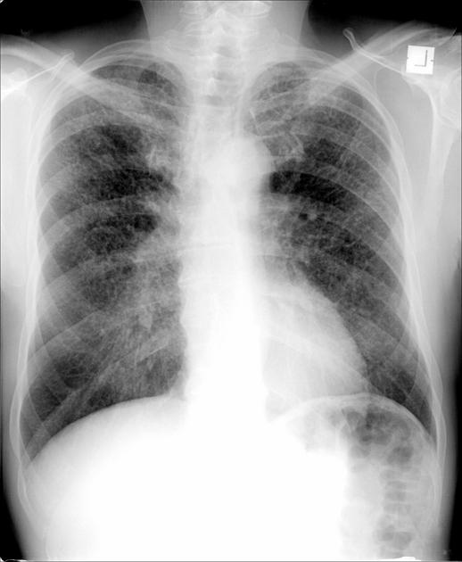
Chest x-ray shows reticular interstitial pattern with preserved lung volume.
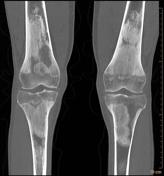

Appearance of symmetrical metaphyseal sclerosis and corresponding increased uptake on Tc-MDP bone scan (shown next).
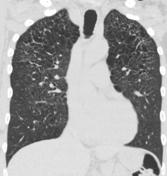
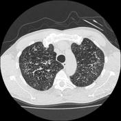
High resolution chest CT reveals predominantly cystic mid and upper zone disease with interstitial thickening, a few nodules, and preservation of lung volumes (the same appearance as Langerhan's cell histiocytosis).
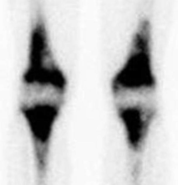
Symmetrical increased uptake on Tc-MDP bone scan corresponding to areas of metaphyseal sclerosis on CT.
Case Discussion
Initial CXR showed reticular interstitial pattern with preserved lung volume. Right knee x-ray revealed femoral and tibial metaphyseal sclerosis. Lung HRCT showed predominantly cystic mid and upper zone disease with interstitial thickening, a few nodules and preservation of lung volumes. Erdheim-Chester Disease (a non-langerhan's cell histiocytosis) was suspected, and the classic symmetrical metaphyseal sclerosis with increased uptake on Tc-MDP bone scan was confirmed when both knees were imaged. The patient died 8 months following symptom onset.


 Unable to process the form. Check for errors and try again.