
Presentation
6 years old female presenting with right thigh mass and pain.
Patient Data
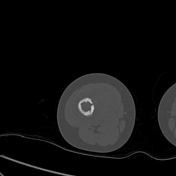

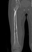

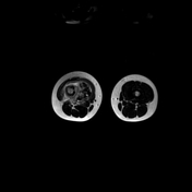
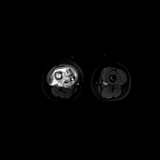
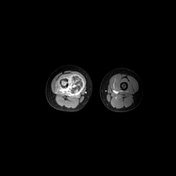
The bone window of this CT scan shows an aggressive type multi-lamellated, elevated and interrupted periosteal reaction.
Endosteal scalloping is also evident and involves most of the femur's diaphysis, but most obvious at the lower third.
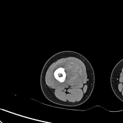
A focal area of frank cortical breach is also seen at the medial side of femur, opposite to which the muscles are displaced away by a heterogenous lobulated mass (better seen on the soft tissue window), hinting significant soft tissue involvement and possible acute hemorrhage.
IMPRESSION: aggressive tumor with significant soft tissue component.
DDx: Ewing sarcoma > osteosarcoma >> osteomyelitis > metastasis.
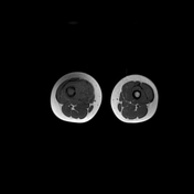
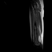
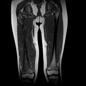
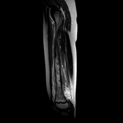
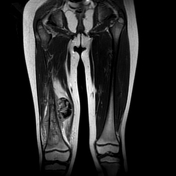
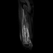
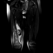
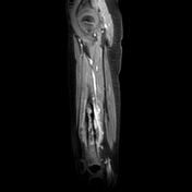


Enhanced MRI of the region shows the true extent of the tumor, which involves most of the marrow cavity of the femur. The associated large soft tissue mass shows heterogenous mainly preipheral enhancement due to internal necrosis. A large acute bleed into the mass is also seen.
Case Discussion
Ewing sarcoma is an aggressive malignant bone tumor seen in unlucky young subjects.
The multi-lamellated periosteal reaction (aka Onion skinning) is a hallmark of this tumor, though, it is not pathognomonic.
Diaphyseal origin of the tumor and associated large soft tissue mass and / or swelling are characteristic features of Ewing sarcoma.
For this case, tissue biopsy confirmed Ewing sarcoma.


 Unable to process the form. Check for errors and try again.