
Presentation
Patient fractured the left clavicle, four months later a suspicious lesion was found on the control radiograph.
Patient Data

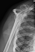
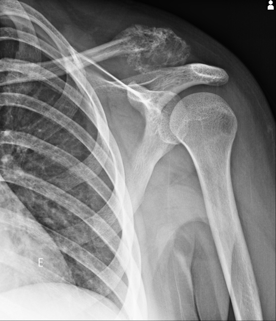
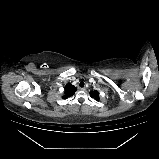
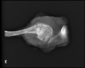
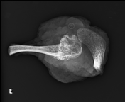
The radiograph shows an expansive lesion in the left clavicle with a permeative appearance. Zanca view demonstrates the difference with the normal clavicle.
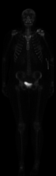

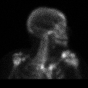
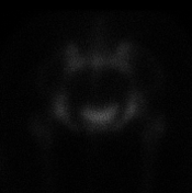

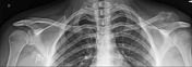
Scintigraphy shows moderate increased uptake at the humeral end of the left clavicle. No signs of bone metastases are observed.
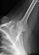
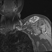
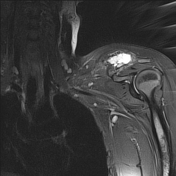
Osteolytic lesion with irregular edges on the left clavicle extending into the supraspinatus muscles, with a suspicious appearance.
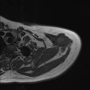
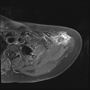
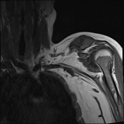
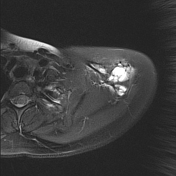
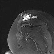

MRI shows a lesion in the left clavicle, with hyposignal on T1, hypersignal on T2, with contrast uptake, compromising the entire distal portion of the left clavicle, acromioclavicular joint, part of the acromion and extending to adjacent soft tissues.

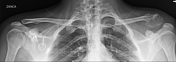
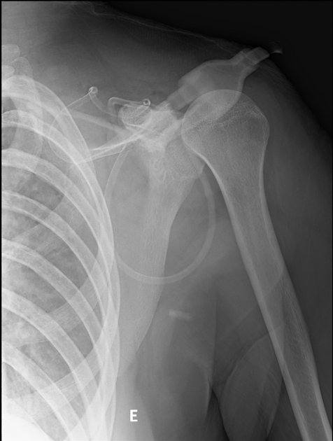
Radiographs taken after surgical resection. The expansive lesion with a permeative appearance.
Case Discussion
The patient suffered a fracture of the left clavicle after a fall from his own height. Four months later, a follow-up X-ray was performed, which revealed an expansile lesion with a permeative appearance, suspicious for neoplasia. Immunohistochemistry showed a pattern compatible with Ewing's sarcoma. Surgical resection was performed after chemotherapy, and histology confirmed the diagnosis.


 Unable to process the form. Check for errors and try again.