
Presentation
Pelvic pain and backache. Known thalassemia major, with history of repeated blood transfusions. Pelvic ultrasound revealed a pelvic mass, for which further imaging was undertaken.
Patient Data


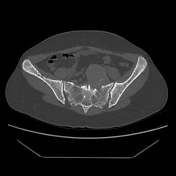
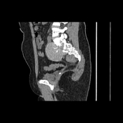
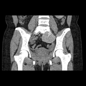
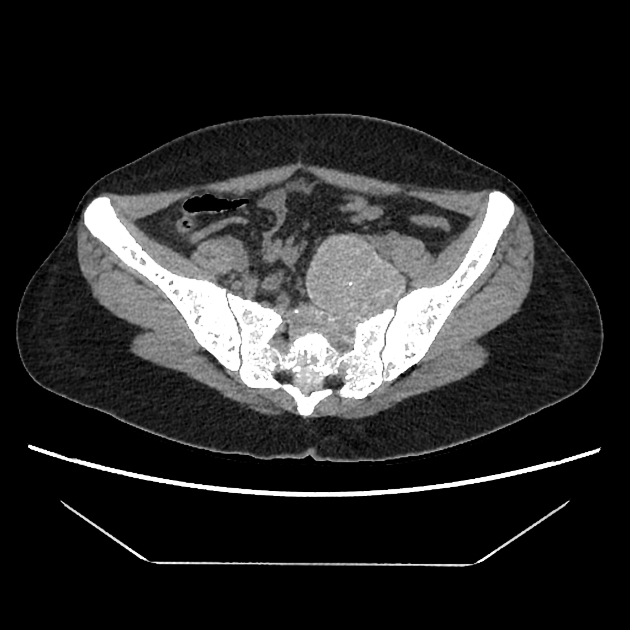
A large well-defined, presacral, inhomogeneous, multilobulated, soft tissue mass, arising from the anterior aspect of upper sacrum is identified. It displays increased attenuation (HU range : +70 to +95). Foci of calcification are seen within the lesion. Similar but smaller lobulated mass is also seen in the anterior part of lower sacrum. Osteopenia and accentuated trabecular pattern of the sacrum, without osseous destruction, is noted in the bone window images.

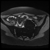
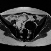
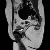
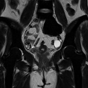
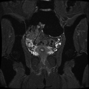
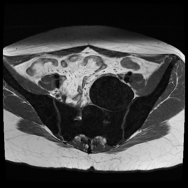
Pelvic MRI shows a large, inhomogeneous, multilobulated, presacral mass, depicting markedly low T1 and T2 signal. The visualized skeleton also shows markedly low T1 and T2 signal.
Case Discussion
In this patient, a known case of thalassemia major, the lobulated presacral mass 3 arising from sacral bone appears to be due to extramedullary hematopoiesis. It typically occurs in paraspinal location 1 (as in this case). Lesion shows attenuation in CT greater than that of a skeletal muscle, and markedly low signal on MRI on both T1-weighted and T2-weighted images, owing to massive iron deposition 1, a sequela of repeated blood transfusions.
In the differential diagnosis of soft-tissue retroperitoneal masses, the low signal intensity on T2- weighted images is in favor of extramedullary hematopoiesis 2.
Very low signal on both T1-weighted and T2-weighted images of visualized skeleton is also a consequence of massive bone marrow iron deposition.


 Unable to process the form. Check for errors and try again.