
Presentation
Fall from height
Patient Data
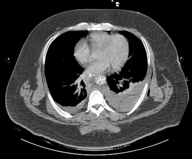
Total body CT; chest scanned at arterial phase and delayed phase (shown).
Deflated balloon catheter in aorta (right femoral approach), its tip at proximal end of descending aorta. Well-placed endotracheal tube and nasogastric tube. Pleural drain on each side, end-holes blocked, resultant soft tissue emphysema.
Small bilateral pneumothorax (seen on lung window). Bilateral pleural effusion (hemothorax), small amount on right and moderate on left, with passive subpleural atelectasis at lung bases.
Extrapleural fat stripe visible on left, separating pleural and extrapleural hematoma as well (extrapleural fat sign).
Thoracic aorta preserved.
Periaortic hematoma extending from T2 to T9. Within it, left paravertebral focus of contrast material extravasation - arterial hemorrhage, perhaps from vertebral artery.
Left clavicular fracture, lateral third.
Burst fracture of T3 and T4 vertebrae, retropulsion at T4 level, exerting pressure on thecal sac. Fracture of left transverse process of T3.
Fracture of right ribs 1, 2, 4, 5, and 8; left ribs 5-7.
Hematoma in intrinsic muscles of back, extending between T2-T4, with contrast material extravasation not seen on arterial phase: venous hemorrhage most probably.
Small amount of free intraeritoneal fluid, likely from diagnostic peritoneal lavage (DPL) done immediately prior to scan.
Case Discussion
This case demonstrates the extrapleural fat sign:
The extrapleural fat stripe can be seen on the left, separating between the intrapleural and extrapleural hematomas.


 Unable to process the form. Check for errors and try again.