
Presentation
Dyspnoea following central venous line placement in the right subclavian.
Patient Data
Age: 70 years
Gender: Female
Download
Info
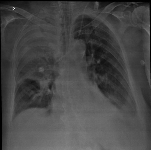
Frontal x-ray shows a large opacity of the right superior pulmonary field, without significant mediastinal shift. A left jugular central venous line is seen. Clinical information was crucial since it was found out that a right side subclavian venous line had failed to pass in multiple attempts.


Download
Info
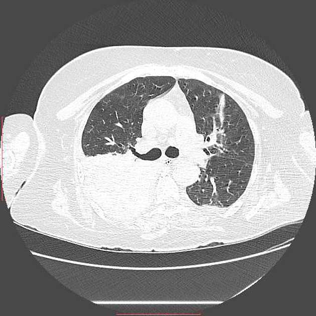
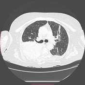
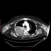
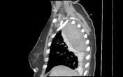
A large collection located in the right superior extrapleural space is seen, with its density suggesting haematic content. A thin line between the large haematoma and the parietal pleura is noticed (extrapleural fat sign).


 Unable to process the form. Check for errors and try again.