
Presentation
Progressive hearing impairment of the left ear with intense tinnitus.
Patient Data
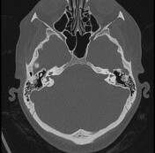

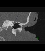

The region of the left facial nerve geniculate ganglion is relatively enlarged may be seen expanded to be correlated with MRI findings.

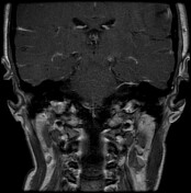
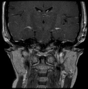
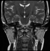
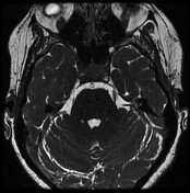

Abnormal soft tissue minute nodular area is seen within the geniculate ganglion of the left facial nerve. It appears isointense on T1, T2, and FIESTA WIs. but shows focal small intense enhancement in the post-contrast study. It measures 0.53 x 0.42 cm in its maximum two dimensions.
Case Discussion
Geniculate ganglion Facial nerve schwannoma is not a common tumor. Among tumors arising from geniculate ganglion, facial nerves are the second most common after hemangioma. The most common symptom is facial paralysis. The DD include: meningioma, dural metastasis, epidermoid, hemangioma of geniculate ganglion. The key diagnostic features of schwannoma include: benign remodeling and widening of geniculate ganglion on CT, avid enhancing lesion.


 Unable to process the form. Check for errors and try again.