
Presentation
Pain right temporal region, prior history of right parotidectomy.
Patient Data
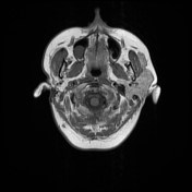

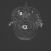
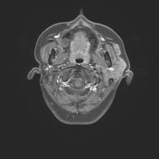

The right parotid gland is not visualized.
Soft tissue is seen anterior to a hypertrophied left parotid that is similar in intensity and enhancement characteristics to the normal parotid parenchyma. No nodules or masses.
Mild edema in left ramus of mandible due to recent dental procedure.
Case Discussion
There are two types of anterior gland extensions:
- facial process: attached to the main gland
- detached glandular mass / accessory parotid gland: mean distance of separation from the gland of 6 mm
Accessory parotid glands are seen in 20% of the population. It usually occurs in a typical location i.e. overlying the masseter muscle and anterior to the parent gland. It lies superficial to the Stensen's duct and drains via an accessory duct into it. They have an independent blood supply from the transverse facial artery. These glands are usually small in size, may be multiple. Tumors of accessory glands are rare.


 Unable to process the form. Check for errors and try again.