
Presentation
Progressive deterioration of cognitive and motor functions.
Patient Data
Age: 14 years
Gender: Male
From the case:
Fahr disease: juvenile onset
Download
Info
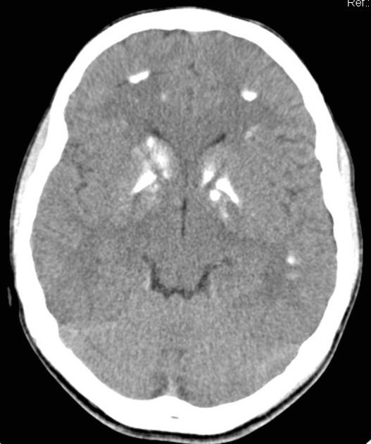
CT imaging reveals bilateral, symmetrical, hyperdense lesions, suggestive of calcifications involving globi pallidi, putamina, caudate nuclei, internal capsules, thalami, dentate nuclei, cerebellum and subcortical white matter.
Case Discussion
Blood chemistry revealed normal serum levels of calcium, phosphorus and alkaline phosphatase.
The CT scan findings when correlated with typical clinical history and normal blood chemistry were suggestive of Fahr disease.


 Unable to process the form. Check for errors and try again.