Presentation
Headache, seizure, right side body weakness and short memory for few years
Patient Data
Age: 30 Years
Gender: Male
From the case:
Fahr syndrome








Download
Info
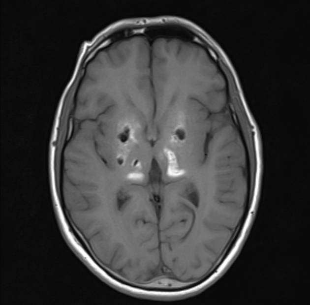
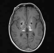
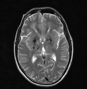
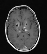
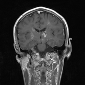
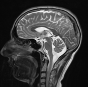
- Significant symmetrical abnormal focal signals in the bilateral basal ganglia, thalami, corona radiata, subcortical white matter as well as dentate neuclei and midbrain, returning T1 hyperintense signal with central low signal, T2 low signal and significant drop out signals in GRE images (representing calcification).
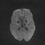
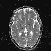
- Nor diffusion restriction neither contrast enhancement.
Case Discussion
The distribution pattern of the calcifications (confirmed on CT, not shown) is characteristic for Fahr's syndrome.


 Unable to process the form. Check for errors and try again.