
Presentation
Finding on bone scan in patient with prior breast cancer.
Patient Data

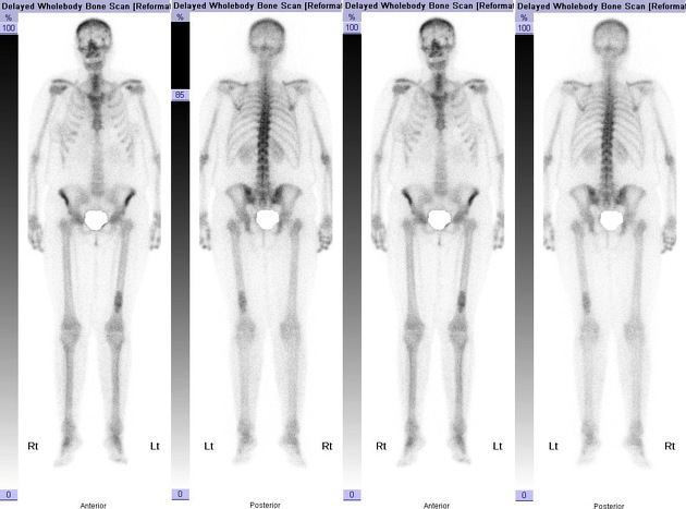
Avid tracer uptake in the distal third of the left femoral diaphysis corresponding with the lesion on plain radiograph.
No other bone lesion.

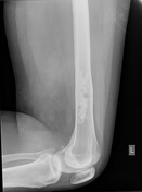
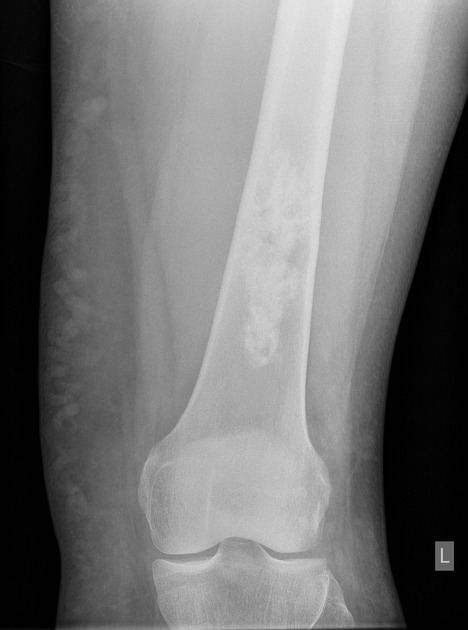
Sclerotic non-expansile bone lesion with a narrow zone of transition in the femoral diaphysis.
Small amount of associated cortical thickening, but no cortical destruction.
No other bone lesion.
Case Discussion
Enchondromas are a relatively common benign medullary cartilaginous neoplasm with benign imaging features. They are often an incidental finding, but can cause potential diagnostic problems in patients with other prior medical history, in particular malignancy.
Increased uptake on the bone scan can be seen with enchondromas.
Plain films correlate with bone isotope scan images is important in the diagnostic process.


 Unable to process the form. Check for errors and try again.