
Presentation
Undisclosed.
Patient Data


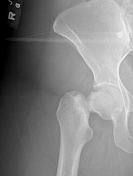
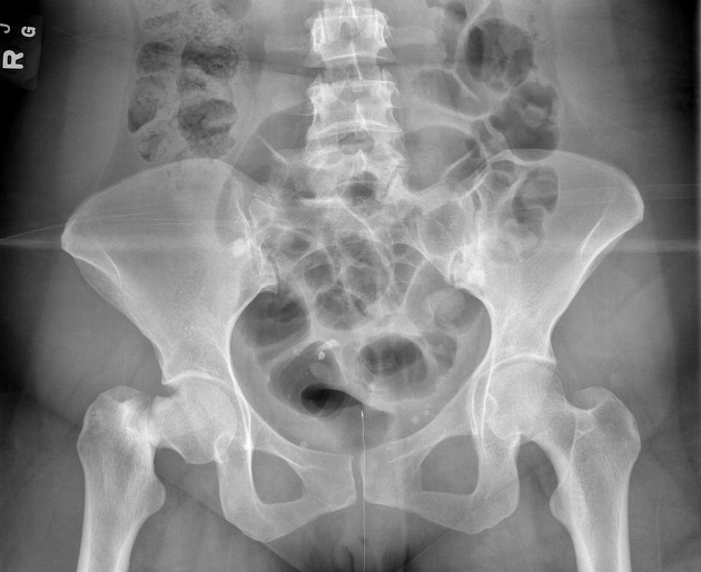
There is a transcervical (bordering on basicervical) fracture of the right femoral neck with resultant coxa vara deformity. This fracture appears non-comminuted. Two dense calcific structures are observed in the pelvic basin to the right of mid-line which are vaguely tooth-shaped. Two 1 cm diameter sclerotic lesions are noted in the right and left ilium bordering the sacroiliac joints, most likely representing bone islands.
The right L5 trasverse process is large and spatulated, and creates a pseudoarticulation with the right sacral ala. At this site an obliquely oriented lucent line with corticated margins is also noted, possibly creating another non-articular motion segment. The left L5/S1 facet joint is moderately hypertrophied and sclerotic indicating facet arthrosis, likely related to the contralateral pseudoarticulation.
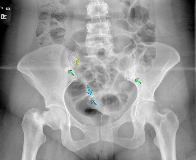
The tooth-shaped calcific structures are indicated by the blue arrows. The bone islands are indicated by the green arrows. The articulating left L5 transverse process is indicated by the yellow arrow.
Case Discussion
The ovarian dermoid cyst was confirmed on a subsequent pelvic MR.
This case is useful to train satisfaction of search, as one may be satisfied with identifying and describing the femoral neck fracture and overlook the multiple other findings.


 Unable to process the form. Check for errors and try again.