
Presentation
Fall one month ago. Ongoing pain left hip.
Patient Data
Age: 80 years
Gender: Female
From the case:
Femoral neck fracture - subacute


Download
Info

Sclerotic line with lateral cortex disruption of the neck of femur in the subcapital region.
From the case:
Femoral neck fracture - subacute


Download
Info
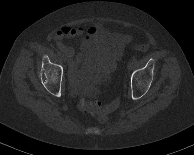
CT confirms a left subacute subcapital neck of femur fracture that is mildly impacted.
From the case:
Femoral neck fracture - subacute
Download
Info

Image intensifier demonstrates the final position of dynamic hip screw and detorsion screw.
Case Discussion
The patient presented to the emergency department at the time of injury but the fracture was not demonstrated on the x-ray or the subsequent CT (even in retrospect a definite fracture line was not seen). An MRI was not performed. Repeat x-rays four weeks after the initial injury show the fracture clearly - the patient proceeded to ORIF.


 Unable to process the form. Check for errors and try again.