
Presentation
Past medical history of polysubstance abuse presenting with altered mental status.
Patient Data


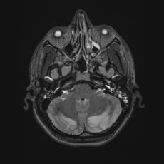
MRI of the brain demonstrates diffusion restriction and T2 hyperintensities involving the cerebral white matter, cerebellar hemispheres, and globus pallidi. 2D chemical shift imaging proton MR spectroscopy shows reduced NAA (N-acetylaspartic acid) in the bilateral basal ganglia, suggesting neuronal loss or dysfunction in this region.
Case Discussion
This case demonstrates restricted diffusion and T2 hyperintensities in the bilateral global pallidi and cerebellar hemispheres as well as involvement of the subcortical white matter (particularly of the motor cortex) and deep white matter of the cerebral hemispheres. These findings are in keeping with a hypoxic-ischemic injury.
In this instance, there is also collateral history from the patient's girlfriend of recent fentanyl inhalation.
Note, the bilateral cerebellar cortical involvement is not the pattern of toxic leukoencephalopathy, seen as a complication of opioid (usually heroin) inhalation (chasing the dragon).
Key learning point:
- An MRI with bilateral, symmetric lesions should raise suspicion for hypoxic, toxic, or metabolic etiology.


 Unable to process the form. Check for errors and try again.