
Presentation
Mother noticed a lump in the right side of the neck with a history of a complicated vaginal delivery.
Patient Data
Age: 3 weeks
Gender: Female
From the case:
Fibromatosis colli

Download
Info
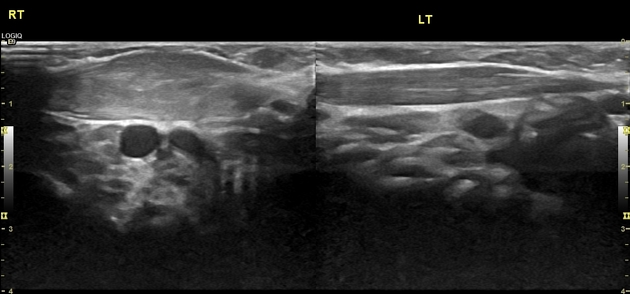
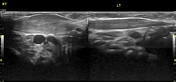
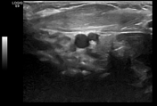
The right sternocleidomastoid muscle shows a diffusely fusiform thickening and shortening.
The left sternocleidomastoid is unremarkable.
Case Discussion
Fibromatosis colli typically presents with a neck swelling at 1-2 months after birth, most commonly as a result of a complicated vaginal delivery and always came with head tilt to the affected site and neck mass.
Ultrasound imaging is the imaging modality of choice in diagnosing fibromatosis colli.


 Unable to process the form. Check for errors and try again.