
Presentation
Incidental finding.
Patient Data
Age: 90 years
Gender: Male
From the case:
Fibrothorax
Download
Info
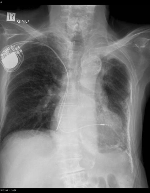
Chest x-ray demonstrates the left-sided chest cavity to be volume depleted with thick pleural calcification in the lower half of the chest. Features are consistent with a fibrothorax. There is no evidence of previous thoracotomy or rib fractures.
Case Discussion
In the absence of prior trauma or surgery the differential includes:
- previous hemothorax
- previous empyema (bacterial or mycobacterial)
The cause of this finding in this individual has been lost to the sands of time.


 Unable to process the form. Check for errors and try again.